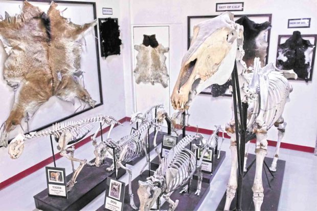
SKIN AND BONES Different sets of skeletons and hides of wild and domestic animals are among the displays that help visitors and students learn more about these creatures. On the museum’s walls and nooks are taxidermied specimens (top), like a monitor lizard and a cattle head. —PHOTOS BY CHRIS QUINTANA AND KIMMY BARAOIDAN
LOS BAÑOS, Laguna, Philippines — The mounted skeletons and hides, and fetuses of animals in glass jars may seem discomforting, but their display has clear learning goals at Dr. Jose A. Solis Museum of Veterinary Anatomy in the University of the Philippines Los Baños (UPLB) here.
“Children should be aware of the importance of our wildlife heritage. And even without seeing [the animals] themselves, if they see bones or pictures, then they would be able to appreciate them,” says Ceferino Maala, 70, a UPLB professor of veterinary medicine and museum curator.
“How many children can go to Palawan to see a peacock or a crocodile or [see] a tamaraw [in Mindoro]?” he asks.
At the museum, which reopened recently, stuffed birds are perched on glass cabinets, animal fetuses float in containers filled with formalin, and skulls of large animals are color-coded inside a large room at UPLB’s College of Veterinary Medicine (CVM).
The exhibits aim not only to educate and entertain visitors but also to promote animal conservation.
PIG FETUS The preserved fetus of conjoined pigs draws the attention of museum visitors. —CHRIS QUINTANA/CONTRIBUTOR
Moreover, the repository of wildlife specimens hopes to encourage students to pursue veterinary medicine. After all, Maala says, “anatomy is the foundation of all medical professions.”
From Diliman to Los Baños
Many of the specimens, like the skeletons and certain bones, were prepared by the late Dr. Jose A. Solis, one of the country’s esteemed veterinary medicine professors. These were part of a collection at the CVM, which was located then at UP Diliman.
The college, along with its Anatomy Collection Center, was transferred to UPLB in 1984.
The center was renovated in 2009 by the VKV VLV Foundation Inc. to commemorate its 50th founding anniversary, and was renamed the Dr. Jose A. Solis Museum of Veterinary Medicine. It underwent another makeover early this year.
Near the museum door, a cow’s head protrudes to greet visitors. Skeletons of a dog, pig, sheep, goat, tiger, cow, horse, false killer whale (Pseudorca crassidens) and other animals of varying sizes are mounted in one area, while animal hides are stretched and hung on the walls.
Some of the specimens can be touched and held by visitors, like the fox skins and large animal skulls, each with a color code to correspond to a specific bone, according to Maala.
Animals with anomalies
The museum also has a small collection of animals with anomalies or malformations, which include conjoined pigs, those with cyclopia (having only one eye or a partially divided eye), puppies with umbilical hernia, and water babies, which are characterized by edema, or an accumulation of excess fluids.
These anomalies are also essential in studying anatomy, Maala says. “In veterinary practice, [those malformations] are also very important, even in human [medicine]. These are reference materials for instruction and for research.”
These are included in the curriculum for veterinary medicine, he says. Students and veterinarians will know, for example, what the offspring may look like if a pregnant animal ingests a certain plant.
Growing collection
Through the years, the museum’s specimen collection has grown from donations given mostly by alumni and a few local institutions.
‘TAMARAW’ SKULL Dr. Ceferino Maala, professor of veterinary medicine and curator of Dr. Jose A. Solis Museum of Veterinary Anatomy in UP Los Baños, shows a skull of a young “tamaraw.” —CHRIS QUINTANA/CONTRIBUTOR
“Recently, [we] got ostrich eggs and skin, and fox skin.” Maala says. A graduate from the Middle East, he adds, donated a mounted skeleton of a gyrfalcon (Falco rusticolus), and a few weeks ago, the skull of a Cambodian crocodile arrived.
A mounted skeleton of a Philippine eagle from the Davao-based Philippine Eagle Foundation is expected in late August.
Maala hopes to expand the museum to an adjacent room, where an audiovisual section could be set up to show photographs and videos of domestic animals giving birth, meat inspection, embalming of animals, stuffing of birds and surgical procedures.
Asked about his dream specimens, Maala says he wants to have a mounted skeleton of a giraffe to serve as museum centerpiece, and a zebra hide.
“When I was in Japan, one professor there said that we could not stop animals from becoming extinct, so what do we do? We put up a museum,” Maala says.
FURRY GIFT This gray fox fur from the United States is a gift to the museum. —CHRIS QUINTANA/CONTRIBUTOR
“Start now to preserve videos, pictures,” he says. “At least you can show to future generations that during this time, there were animals like these roaming the surface of the earth.”